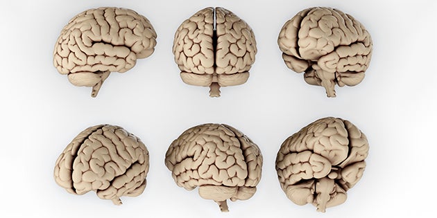

Computer science professor Brent Munsell has helped develop a new technique for diagnosing autism using images of the brain. The red and blue nodes represent different regions in the brain such as visual processing, face and word recognition and self awareness. The yellow lines map the connections between them.
Despite all the marvels of modern medicine, the human body still holds a lot of secrets. The brain, in particular, remains a mysterious network of chemical and electrical pathways that control everything from movement to memory to emotion.
But College of Charleston computer science professor Brent Munsell is one step closer to unraveling some of the brain’s biggest enigmas – mainly what causes certain neurological disorders (think autism, Alzheimer’s or Parkinson’s).
Munsell, along with researchers at the departments of Psychiatry and Radiology at the University of North Carolina Chapel Hill and the School of Software at Tsinghua University in Beijing, China, has developed a new method for analyzing neuroimaging data in an effort to better diagnose impaired brain function.
Many neurological disorders – or malfunctions of the brain – are still largely diagnoses of exclusion rather than ones based on detectable, scientific evidence. Often illnesses such as autism or Parkinson’s are diagnosed only after a doctor has ruled out other possible conditions. And that’s what Munsell and his research partners set out to change by developing a better computational technique that more comprehensively maps the different sections of the brain and how they’re connected.

Professor Brent Munsell
Using existing data from a study of children and adolescents at the University of California, Los Angeles, the group of researchers used a new approach of combining functional and structural elements of the brain (the parts of the brain that control visual, motor skills, etc. and the wiring that connects each part of the brain) to identify abnormalities – which in this case indicated a high risk for autism.
The new approach yielded exceedingly promising results. Images using the new technique, Munsell says, yielded an 88 percent recognition rate for autism compared to the 60-70 percent available through current state-of-the-art technology.
“This is a really meaningful thing,” Munsell says of the results. “This approach to brain imaging gives us a look at how the brain is organized (functionally and structurally). Based on those relationships, we can determine if you have autism or not.”
Just how meaningful are these findings? Well to start with, the Medical Image Computing and Computer Assisted Intervention Society will publish the research in its medical publication. Munsell and his colleagues in North Carolina and China will also travel to Greece in October 2016 to present their findings at the society’s annual international conference.
Under the direction of Munsell, the Department of Computer Science at CofC has been leading a variety of research efforts in this field in its Machine Learning and Medical Image Analysis Lab, analyzing similar data from epilepsy, Alzheimer’s and traumatic brain injury studies.
This latest effort for evaluating autism patients, Munsell says, has the potential to reach well beyond just one condition. Medical professionals could also use it to more definitively diagnose other brain disorders such as Alzheimer’s disease and Parkinson’s disease.
“The ultimate goal is to improve health care in general,” Munsell says. “The more accurately a doctor can give a diagnosis, the better.”


